Friday, October 30, 2009

Sialoliths


I decided to do my blog for this week on salivary gland stones since this is a common reason for soft tissue neck scans in our facility. I became interested in this after we scanned a young female who had a past history of salivary gland stones. She presented with pain and swelling in her left jaw. She told me a previous stone had been removed in her dentist’s office. The dentist used a small tweezers to remove it from the duct opening. The scan this time showed a very large stone in her duct. I wish I had an image but I can’t remember the patient’s name or when we scanned her. Anyhow, I doubt this stone was going to be removed easily!

Salivary gland stones are most common in the submandibular gland duct (75 - 80%) with the rest usually found in the parotid gland duct (25%). Stones are rare in the sublingual gland ducts. The stones are usually made up of calcium and can range in size from less than 1 mm to several centimeters. They are more common in males, after age 40, and some people are just more prone to developing them (just like some people are more prone to developing kidney stones). It is thought that stones form more commonly in the submandibular glands because the saliva found in these glands is thicker and has to drain uphill.

The symptoms of a salivary gland stone are pain and swelling in the jaw or face, or swelling of the gland itself. Another symptom is dry mouth. The symptoms are all more common at mealtimes. This is because the salivary glands produce extra saliva when you eat and the saliva doesn’t have anywhere to go because it is blocked by the stone. Infection and abscess can occur if the stone isn’t removed. Diagnosis can be by plain film x-ray, sialography, or CT.

Small stones at the duct opening can often be removed by a dentist. Endoscopy can be used to capture and remove stones using a dialator, basket or balloon. Open surgery is the last option. If stones recur in just one gland the individual gland can be removed and the mouth will still make enough saliva.

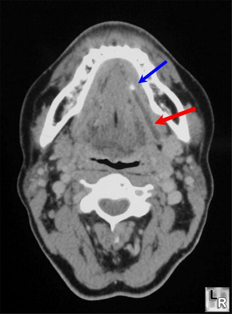
This CT image shows a stone in the submandibular duct (blue) and the dialated duct (red).

Image:

http://www.learningradiology.com/archives04/COW%20096-Sialolithiasis/sialolithcorrect.htm

References:

http://health.nytimes.com/health/guides/disease/salivary-duct-stones/overview.html

http://othn.iusm.iu.edu/salivaryglandendoscopy/

http://www.patient.co.uk/health/Salivary-Gland-Stones.htm

Tuesday, October 27, 2009

Vein of Galen Malformation

Vein of Galen Malformation

This malformation can sometimes be diagnosed with prenatal ultrasound but most commonly is detected when a newborn infant develops rapid heart failure. Other symptoms are hydrocephalus, developmental delay or seizure. The malformation is caused by a blood vessel in the brain that is present during embryonic development but supposed to disappear before the baby’s birth. Because the vessel persists in the brain it allows for a direct connection between the arterial and venous systems of the brain. So, rather than flowing through capillaries as it is supposed to the blood flows directly between the two systems. Because arteries flow faster than veins are capable of draining, this connection puts a great strain on the organs of the body, most notably the heart. That is another reason for the hydrocephalus – the brain cannot drain the blood fast enough and it builds up in the brain. The malformation needs to be treated to prevent continued symptoms and death. The old method (not very effective) of treating the problem was surgery, but today embolization is more commonly used and has a higher success rate. The purpose of embolization is to reduce the blood flow that is feeding the malformation. The malformation is gradually blocked off using either coils or glue which is introduced through a femoral catheter. Sometimes it takes more than one embolization procedure to treat the malformation. Prognosis is usually good if the disorder is caught and treated before developmental delays develop. I was surprised to find a support group on the web!

Reference:

Vein of Galen Malformation Support Group. Accessed October 27, 2009 at http://www.veinofgalen.org.nz/about_vein_of_galen/signs,_symptoms_and_diagnosis

Image:

This image is a sagittal MIP from a CT cerebral angiogram on an infant and shows a Vein of Galen malformation. The posterior superior sinus, transverse and sigmoid sinuses are enlarged.

Image from: Radiopaedia.org, Accessed on October 27, 2009 at http://radiopaedia.org/cases/vein-of-galen-tof-mrv

Monday, October 12, 2009

Fungal Sinusitis

Chronic Sinusitis caused by fungus

In 1999 researchers at the University of Buffalo showed that fungal organisms were found in the mucus of 96% of patients who had sinus surgery for chronic sinusitis. In addition, inflammatory cells were found to be clumped around the fungi. This led the researchers to test the theory that the chronic sinusitis was in actuality an immune response to the fungus. Their research found that most people have airborne fungi lodged in the mucus lining of their sinuses but only people prone to chronic sinusitis have an immune reaction to the fungi. When the fungi are attacked it can cause the following immune responses in the sinuses; long term nasal congestion, thick mucus, chronic headache, bacterial infection and loss of smell. Chronic sinus headache can have a serious impact on quality of life.

Researchers at the Mayo Clinic and University of Buffalo conducted a study in 2004 using the fungicide Amphotericin-B applied intranasally and found that the “treatment group showed a significant decrease in the inflammatory thickening of the sinus membranes compared to the control group. “ Also, 70% of the patients showed a decrease in the amount of nasal swelling vs. the control group when viewed by endoscope. The study subjects were followed with CT scans of the sinuses at study start and again at six months, as well as endoscope at start of study, three and six months. CT results indicated an 8.8% decrease in mucus thickening in the treatment group vs. an increase of 2.5% in the placebo group.

The researchers hope this study will lead to the development of a fungal product for the treatment of chronic sinusitis. I wasn’t able to find any information on an FDA approved fungal product for this use so testing must still be on-going. (Note: Mayo Clinic sold the rights to the drug to Accentia Biopharmaceuticals and this company was granted FastTrack FDA status for SinuNase in 2006. SinuNase is an intranasal Amphotericin B formulation. However, Accentia is a biotech company that was delisted from the Nasdaq in 2008 and appears to be having financial problems, so future marketing may be uncertain).

I just thought this was an interesting article because most people think sinusitis is only bacterial and this suggested another possible cause. The CT images show the same patient pre and post study after treatment with Amphotericin B. You can see the improvement in the right maxillary sinus in the image on the right. I would imagine that the patient was feeling much better post study because they had a lot of mucus and swelling - ouch!

Reference and images source:

Baker, L. (2004). Chronic sinusitis caused by fungus. UB Reporter, Archives (April 1, 2004). Accessed October 12, 2009 at http://www.buffalo.edu/ubreporter/archives/vol35/vol35n28/articles/Sinusitis.html

Accentia granted FastTrack Status for SinuNase. Accessed October 12, 2009 at http://www.encyclopedia.com/doc/1G1-144391918.html