Friday, April 16, 2010

Poor King Tut


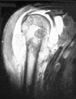
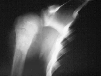
Poor King Tut. CT images of his body indicate that he may have died of a broken leg. When his body was x-rayed in 1968 results showed he had a broken skull so scientists of the time believed he died a violent death. As imaging became more advanced scientists decided to CT his body. One advantage of this method was the entire body could be imaged without having to move it. 1,700 images were acquired using CT in 2006 (see reconstructed image on left). The CT results indicated no skull trauma but did show skull damage most likely due to the mummification process. This CT image of King Tut’s left knee indicates a distal femur fracture. The arrow points to dense embalming material around the fracture which indicates that it occurred shortly before his death. Numerous other smaller leg fractures were also found, leading scientists to theorize that the King broke his leg badly shortly before his death. Because the fracture was open it became infected and killed him. The addition of DNA testing to the CT results (published in 2010) revealed that King Tut’s parents were most likely sister and brother. In addition to being the product of siblings, King Tut also had bone disease and malaria, both which would have further weakened his immune system so he was unable to fight off the infection caused by his broken leg. King Tut also had club feet and a cleft palate. The club feet would have explained the presence of the 130 walking canes in his tomb. I thought this was an interesting knee pathology for the week – certainly something different! It shows how current imaging technology in combination with other modern tests can help to solve mysteries of the past.

Source and Image:

Hayes, J. (2006). King Tut’s death official: broken leg. Accessed April 16, 2010 at Cosmos online, at http://www.cosmosmagazine.com/news/882/king-tuts-death-official-broken-leg

Discovery Channel. Discovery News. Accessed April 16, 2010 at http://dsc.discovery.com/news/briefs/20050307/gallery/tutscan_zoom.jpg

Hasan, L. & Phend, C. (2010). How King Tut Died Revealed in New Study. ABC News Feb 16. Accessed April 16, 2010 at http://abcnews.go.com/Health/LivingLonger/king-tut-died-revealed-study/story?id=9853119&page=1

Wednesday, March 17, 2010

Osteosarcoma


Osteosarcoma is a cancer that originates in the bones and is the most common type of malignant bone tumor. It is not a cancer that has metastasized to the bones from another location in the body. It is most prevalent at the metaphysis of the growth plate in the long skeletal bones and is found most often around the knee. The second most typical presentation is in the proximal humerus near the shoulder area. Teenagers are the largest affected group. About 400 of the 900 new cases in the United States each year are in teenagers. The risk of osteosarcoma is thought to be higher in teenagers because they are growing rapidly. Boys tend to get this type of cancer more frequently than girls. Symptoms include bone pain or swelling which worsens with activity. The first imaging study ordered is usually plain film x-ray. Diagnosis is confirmed with needle biopsy, lab and blood tests. CT, MR, Nuclear Medicine and PET scans are useful for surgery planning and staging. Tumors are considered either “localized” or “metastatic” and are staged accordingly. The 5-year survival rate for localized osteosarcoma is 60% to 80%. For metastasized osteosarcoma the rate is 15% to 30% for 5 years. Treatment has improved since the 1960’s when the most common treatment was amputation of the affected limb. Treatment today involves chemotherapy pre and post surgery. Once the tumor is shrunk using chemotherapy it is then removed. Patients whose tumors respond well to pre-surgery chemotherapy usually have a better long term prognosis. Limb sparing surgery is used whenever possible. Bone grafts or metal rods can be used to replace bone in many cases thus sparing the patient an amputation (American).

I thought these were interesting images. They all show the same right shoulder of this teenage boy with osteosarcoma. You can see the outward physical signs of the tumor as well as the results of the imaging studies. The photograph of his chest shows swelling of the deltoid. The plain film x-ray shows the radiodense area of tumor in the bone as well as the soft tissue swelling. The bone scan shows the difference in radionuclide uptake in the involved vs. the uninvolved shoulder. The MR image indicates how much the bone tumor has spread into the soft tissues of the shoulder from the bone.

Reference:

American Cancer Society. Cancer Reference Information. What is Osteosarcoma? Accessed March 17, 2010 at http://www.cancer.org/docroot/CRI/content/CRI_2_2_1X_What_is_osteosarcoma_52.asp?sitearea=

Images:

Mehlman, C. & Cripe, C. (2008). Osteosarcoma. Accessed March 17, 2010 at http://emedicine.medscape.com/article/1256857-overview


Monday, February 15, 2010

Ovarian Cancer

Ovarian Cancer is the second most common gynecologic cancer and is most common in women aged 40 to 65. Risk factors include early menstruation with late menopause, obesity, and a family history of ovarian, breast or colon cancer. Other risk factors may include using either the fertility drug Clomid or hormone replacement therapy. The American Cancer Society estimates 21,550 new cases of ovarian cancer and 14,600 deaths due to ovarian cancer in 2009 (ACS). Pregnancy, breastfeeding, birth control pills, ovarian removal, and tubal ligation/hysterectomy may offer some protection against ovarian cancer.

Only about 20% of ovarian cancers are found in the early stages and early detection is the best survival tool. Symptoms may include stomach or abdominal swelling/bloating, pelvic pressure, a rapid feeling of fullness after eating, urinary urgency and/or urinary frequency. Other women have complained of symptoms like persistent indigestion, a change in bowel habits, painful intercourse and lower back pain. The problem is that many of these symptoms can also be caused by such things as PMS, irritable bowel syndrome, urinary disorders or other diseases. The key is if these symptoms are persistent, last beyond a few weeks and worsen over time. If this occurs a woman should see her gynecologist for testing.

Ultrasound is usually the first modality used for the diagnosis of ovarian cancer and can determine the presence or absence of a mass with 97% accuracy (GLOWM). CT is often used to determine tumor size, location and degree of metastasis to adjacent lymph nodes or organs. Blood tests are used to determine levels of CA-125, a blood protein often found at higher levels in women with ovarian cancer. During surgery many tissue samples are taken in the abdomen and pelvis to determine cancer staging. Staging ranges from stage I (confined to ovaries) to Stage IV (spread to distant organs such as liver or lungs). Five year survival rates vary from 80% for Stage I to 18% for Stage IV. Treatment consists of surgery, chemotherapy and radiation.

This image shows a bilateral stage IB Ovarian cancer in a 47 year old. Despite the large masses (see O) the cancer is still confined to the ovaries.

Image:

http://www.glowm.com/resources/glowm/graphics/figures/v4/0540/034f.jpg Accessed February 15, 2010.

References:

ACS. American Cancer Society. Overview: What is Ovarian Cancer? Accessed February 15, 2010 at http://www.cancer.org/docroot/CRI/CRI_2_1x.asp?rnav=criov&dt=33

GLOWM. The Global Library of Women’s Medicine. Ovarian Cancer. Accessed February 15, 2010 at http://www.glowm.com/?p=glowm.cml/section_view&articleid=265&SESSID=haaj72nm2tvd91b1f18m3chhk5

MayoClinic.com. Ovarian Cancer. Accessed February 15, 2010 at http://www.mayoclinic.com/health/ovarian-cancer/DS00293/DSECTION=symptoms

National Cancer Institute. Ovarian Cancer. Accessed February 15, 2010 at http://www.cancer.gov/cancertopics/types/ovarian