Osteosarcoma is a cancer that originates in the bones and is the most common type of malignant bone tumor. It is not a cancer that has metastasized to the bones from another location in the body. It is most prevalent at the metaphysis of the growth plate in the long skeletal bones and is found most often around the knee. The second most typical presentation is in the proximal humerus near the shoulder area. Teenagers are the largest affected group. About 400 of the 900 new cases in the United States each year are in teenagers. The risk of osteosarcoma is thought to be higher in teenagers because they are growing rapidly. Boys tend to get this type of cancer more frequently than girls. Symptoms include bone pain or swelling which worsens with activity. The first imaging study ordered is usually plain film x-ray. Diagnosis is confirmed with needle biopsy, lab and blood tests. CT, MR, Nuclear Medicine and PET scans are useful for surgery planning and staging. Tumors are considered either “localized” or “metastatic” and are staged accordingly. The 5-year survival rate for localized osteosarcoma is 60% to 80%. For metastasized osteosarcoma the rate is 15% to 30% for 5 years. Treatment has improved since the 1960’s when the most common treatment was amputation of the affected limb. Treatment today involves chemotherapy pre and post surgery. Once the tumor is shrunk using chemotherapy it is then removed. Patients whose tumors respond well to pre-surgery chemotherapy usually have a better long term prognosis. Limb sparing surgery is used whenever possible. Bone grafts or metal rods can be used to replace bone in many cases thus sparing the patient an amputation (American).
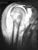
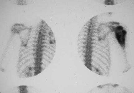
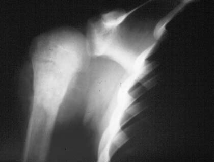
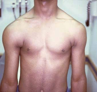
I thought these were interesting images. They all show the same right shoulder of this teenage boy with osteosarcoma. You can see the outward physical signs of the tumor as well as the results of the imaging studies. The photograph of his chest shows swelling of the deltoid. The plain film x-ray shows the radiodense area of tumor in the bone as well as the soft tissue swelling. The bone scan shows the difference in radionuclide uptake in the involved vs. the uninvolved shoulder. The MR image indicates how much the bone tumor has spread into the soft tissues of the shoulder from the bone.
Reference:
American Cancer Society. Cancer Reference Information. What is Osteosarcoma? Accessed March 17, 2010 at http://www.cancer.org/docroot/CRI/content/CRI_2_2_1X_What_is_osteosarcoma_52.asp?sitearea=
Images:
Mehlman, C. & Cripe, C. (2008). Osteosarcoma. Accessed March 17, 2010 at http://emedicine.medscape.com/article/1256857-overview

No comments:
Post a Comment