

For my blog this week I wanted to learn more about Odontoid fracture. The facility where I work weekend option diagnostic x-ray is going to become a Trauma One facility in the next few years so we will be seeing more severe trauma. And we recently had a patient come into my clinical location for repeat scans post C-2 fracture. He had been in a car accident 8 months prior and sustained a severe whiplash injury to his neck. He told me that he had been in the back seat of the car when it was rear-ended at high speed. The fact that he wasn’t wearing his seat belt actually saved his life because he was only flung forward. He was told by his physicians that had he been caught by the seat belt and flung backward as well the force would have severed his spinal cord and killed him instantly.
I was amazed that he walked into the clinic. He told me he was put into a collar and onto a backboard at the accident scene. He had an immediate CT scan in the ER which showed the fracture. His first surgery was an anterior screw for stability similar to image #4 on the right. I forgot to ask him if he wore a halo after this surgery but I would guess that he did since this seems to be the conventional treatment. He told me that the initial screw started to migrate causing him pain and numbness so a second surgery was performed replacing the first screw with posterior hardware. We were scanning in CT that day to assess the healing and stability of the second set of hardware (similar to image #1 on the left). Unfortunately I don’t have his actual images to post here.
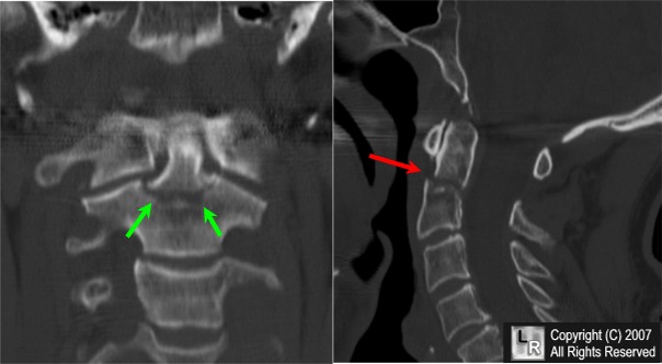
I learned that there are 3 types of Odontoid fracture and most are caused by MVA’s and falls. 1/3 of C-spine injuries occur at C2 and ½ at C6-C7. The majority of fatal injuries occur at C1 or C2 and they tend to occur due to extreme flexion, extension or rotation. They are classified by their location on the dens. Type I goes through the very top of the dens and is very rare, only about 5% of cases. Type II is shown in the middle two images here and is through the base of the dens. This represents about 60% of dens fractures and is the most common type. Type III is through the body of C2 and doesn’t actually involve the dens and is about 30% of fractures. CT is better for demonstrating fractures and MR is used to show the extent of soft tissue, disc, ligament and spinal cord involvement.
Type I fractures are usually treated with a hard collar. Type II fractures are treated surgically as described above with the addition of a stabilizing halo for several months. My patient told me he just feels lucky to be alive. After seeing his images, I must say I agree!
References and Images:
http://www.learningradiology.com/caseofweek/caseoftheweekpix2007-1/cow248arr.jpg
http://www.thebarrow.org/stellent/groups/public/@xinternet_con_bni/documents/webcontent/bqjpg120.jpg

No comments:
Post a Comment